Expansion Microscopy-based imaging for visualization of mitochondria in Drosophila ovarian germline stem cells
- Author:Chi-Hung Lin, Tzu-Yang Lin, Shao-Chun Hsu, Hwei-Jan Hsu
- Journal: FEBS Open Bio12(2022) 2102-2110 https://febs.onlinelibrary.wiley.com/doi/10.1002/2211-5463.13506
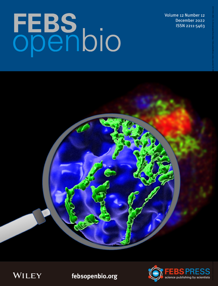
Recent studies have shown that mitochondrial morphology can modulate organelle function and greatly affect stem cell behavior, thus affecting tissue homeostasis. As such, we previously showed that the accumulation of fragmented mitochondria in aged Drosophila ovarian germline stem cells (GSCs) contributes to age-dependent GSC loss. However, standard immunofluorescence methods to examine mitochondrial morphology yield images with insufficient resolution for rigorous analysis, while 3-dimensional electron microscopy examination of mitochondrial morphology is labor intensive and allows only limited sampling of mitochondria. To overcome these issues, we utilized the expansion microscopy technique to expand GSC samples by 4-fold in combination with mitochondrial immunofluorescence labeling. Here, we present a simple, inexpensive method for nanoscale optical imaging of mitochondria in the germline. This protocol may be beneficial for studies that require visualization of mitochondria or other fine subcellular structures in the Drosophila ovary.